


Much of my coaching is spent on those affected by mold. Whether they are living in it, working around it, or have lived in it, it’s a major issue. Mold is also one of the most gaslit topics around, particularly for landlords, colleges, management companies for apartments, or employers.
Most conversations go like this: “Hey, there is a bunch of black mold and water damage in my ceiling, and ever since I was exposed, my health has gone downhill.”
“THAT’S NOT MOLD, THAT’S SOMETHING ELSE, YOU’RE MAKING IT UP! MOLD DOESN’T DO THAT!”
It’s just like when Monsanto execs said they would guzzle down the weedkiller glyphosate. One exec said this in an interview, only for the journalist to challenge him on it. Shockingly, the interview was cut short by the Monsanto exec... Turns out he didn’t want to drink glyphosate.
When someone gaslights you on mold, gladly tell them to take some of that mold home, hose down all their drywall, and then plaster mold all over the walls. In a month, they might change their mind. Reality is, they just don’t want to pay for fixing it.
Even though mold makes up much of my coaching, the more I deal with mold clients, the more I realize how ubiquitous it truly is. Given that 50% of buildings in the US are water damaged, and Americans spend 90% of their time inside, should any of us be shocked by this epidemic?1
In this post, I am going to go over how to approach mold toxicity. I already have two blogs out on what mold does to the body and how to address it in one’s home. Both are fantastic articles in my opinion.
Ritchie Shoemaker
One cannot talk about mold without first talking about Dr. Ritchie Shoemaker, the godfather of biotoxin illness. A Maryland family doctor, Shoemaker first came onto the scene when a wave of his patients mysteriously fell ill after exposure to a toxic microorganism bloom (Pfiesteria piscicida) in the Chesapeake Bay.
After seeing enough of these patients exhibiting similar symptoms, he connected the dots: mapping how biotoxins like Pfiesteria piscicida drive a shared cluster of brutal immune responses in the body.
Eventually, after seeing similar symptom clusters in people living in water-damaged buildings and being exposed to mold, he coined the term Chronic Inflammatory Response Syndrome (CIRS).
CIRS is what happens when a genetically susceptible person meets a biotoxin, and their immune system locks into a chronic state of “fight or flight,” as I like to put it.
Whether the trigger is mycotoxins, cyanobacteria, or Lyme, the body responds the same way: a relentless, self-perpetuating inflammatory response. That common reaction is CIRS.
Rather than testing for the biotoxin directly, which can have some efficacy issues, although I still find that data useful, he proposed measuring the inflammatory response itself.
In his case, when you have enough ‘mystery illnesses’ pop up at your front door, presenting in identical manners, and have current or prior exposures to something like mold, the real gold standard would be measuring how your body is responding to that exposure.
Some people can live in mold and have zero issues whatsoever, while some develop CIRS. It’s like measuring a clue left in the blood, and when all the CIRS blood markers come together, you have your answer.
Here are the markers Dr Shoemaker identified in conjunction with CIRS2:
Alpha-melanocyte-stimulating hormone (α-MSH): is a peptide hormone responsible for quite a bit in the body (see Dr Jack Kruse’s work here; I plan on writing about this down the line). It is cut from the parent protein POMC, which is created in response to UV light (more on this later), and is a small peptide, 13 amino acids to be exact. It helps regulate inflammatory pathways in the body, including NF-κB (the master switch of inflammation), chemokine receptors, which help direct immune cell traffic, production of proinflammatory cytokines and mediators, synthesis of the anti-inflammatory cytokine IL-10, T cell proliferation, expression of antioxidative enzymes, and apoptosis (programmed cell death). It’s even been shown to be important in gut barrier function.3
Transforming Growth Factor Beta-1 (TGF-β1): An immune cytokine responsible for tissue repair, fibrosis, and immune regulation. Under normal circumstances, TGF-β1 keeps the immune system in check; when it runs hot, it promotes something called Th17 dominance and suppresses your regulatory T cells, the commander T cells that prevent friendly fire, leaving the immune system stuck in “soldier mode” and tipping the body toward chronic inflammation and autoimmunity. It’s also the body’s scar-builder, which is why fibrosis, vascular issues, and poor circulation are so common in mold.4
Matrix Metalloproteinase-9 (MMP-9): An enzyme responsible for remodeling tissue by breaking down the extracellular matrix (the scaffolding between cells) and the tight junctions that seal the barrier function of cells. Through excessive stimulation, that breakdown of tissues — blood vessels, gut lining, and tight junctions — contributes to leaky gut and a degraded blood-brain barrier.
Vascular Endothelial Growth Factor (VEGF): A signal protein responsible for the building of new blood vessels through angiogenesis. Proper VEGF levels allow oxygen and nutrients to be effectively perfused throughout tissues. With mold, it usually runs low, resulting in poor blood perfusion.
C4a: A complement fragment fired off by the innate immune system, and one of the first alarms to spike after biotoxin exposure. Chronically high, it’s tied to reduced blood flow at the tissue level, which fuels the fatigue, air hunger, and brain fog.
VIP: A master regulatory neuropeptide governing inflammation, blood flow, and pressure in the lungs. It’s typically depleted in CIRS, the sign the body’s regulatory reserves are running on empty.5
All in all, these markers can tell a pretty clear story about how the immune system is reacting to biotoxins like mycotoxins. Immune dysregulation can be pinned back to a majority of the chronic issues many folks have today, and Shoemaker has even said TGF-β could be a pivotal marker for future autoimmune care.
“I suspect that the next textbook of autoimmunity and rheumatology will be one dedicated to treating high TGF beta-1 and restoring control of T-regulatory cells.”6
I agree, whether it be biotoxins, heavy metal toxicity, glyphosate exposure, SIBO and endotoxemia, or the many plagues of modern living, there is much overlap regarding the effects on the immune system.
Bile and Binding
If you enjoy any of my content, you know I am obsessed with the liver and bile. Bile is one of the main routes our body uses for excreting toxins. Poor bile flow means poor toxin excretion, and lack of bile + lack of binders makes for a recirculation fiasco.
In my heavy metals article, I cited a study that showed 90-95% of bile is reabsorbed in the small bowel, and the heavy metals alongside it. This is a well-known phenomenon, and creates a negative feedback loop in the liver, as toxins continually drain the liver’s antioxidant stores and clog bile.
Shoemaker argues for bile acid sequestrants, where bile is bound and excreted, giving no opportunity to recirculate mycotoxins. He utilizes things like cholestyramine and Welchol to sequester bile into stool.
These drugs were initially used as LDL cholesterol-lowering drugs, as cholesterol is an input to bile, and if it is sequestered, it has no opportunity to be reabsorbed again. You’re pretty much pooping out excess cholesterol.
Same principle applies for the mycotoxins in the bile: the goal is to poop or pee them out, not reabsorb them.
These are the main binders used in the Shoemaker protocol, and they are quite effective, particularly for mycotoxins like OTA. 7
After use of these binders, and MARCoNS are dealt with in the nasal cavity, Shoemaker follows up with VIP nasal peptide, which helps regain immune regulation, and normalize the CIRS markers.
I have much respect for Dr Shoemaker, and am extremely grateful I can build off of his extensive research and experience with the 1000s of mold patients he has had over the years.
It allows for coaches like me, who have their own unique protocols, to build on top of proven strategies.
I personally think there is much more that can be done in the context of mold!
So let’s get into those strategies!
Binders
Everyone interested in health knows what a binder is: most commonly activated charcoal, which binds to toxins in the gut to be excreted via stool.
With binders, there is much nuance to explore.
A binder acts like a magnet; it grabs onto something of the opposite charge. A single binder will not grab onto every single toxin, because certain toxins have certain affinities, and certain binders have certain affinities.
Given there is a diversity of mycotoxins, it means there are different approaches to binding certain mycotoxins.
This is precisely why I like a multi-spectrum binder, and why I think Dr Shade, the founder of Quicksilver Scientific, understands what the heck he is doing with detox. These principles extend to heavy metals, glyphosate, PFAS, plastics, and many of the other environmental toxins we are exposed to, and often have while addressing mold.
So let’s start with the various types of binders and what they suit best.
Activated charcoal
Activated charcoal is a universal toxin binder, found in grocery stores, pharmacies, and even used in the hospital for acute toxicity. It’s a nonspecific binder, meaning it grabs onto a multitude of things, and is derived from burning something like coconut shells, creating a large porous molecule.
Here are some studies on mycotoxins and charcoal:
Deoxynivalenol (DON)/Trichothecenes: Pigs received a single large dose of the mycotoxin DON alone or in combo with activated charcoal. The dose without charcoal showed significant quantifiable plasma amounts of DON, whereas the charcoal group had completely prevented the absorption of DON and no plasma amounts could be detected.8
Deoxynivalenol (DON) & Nivalenol (NIV)/Trichothecenes: Using a simulated digestive tract of pigs, researchers were able to evaluate how well DON and NIV would be absorbed with and without charcoal. Without charcoal, 51% of DON and 21% of NIV were absorbed predominantly in the jejunum (middle part of the small intestine). With 2% charcoal added to the trichothecenes concoction, it reduced DON absorption to 21% and NIV to 12%. The author also notes how charcoal was even more effective at binding zearalenone in prior research.9
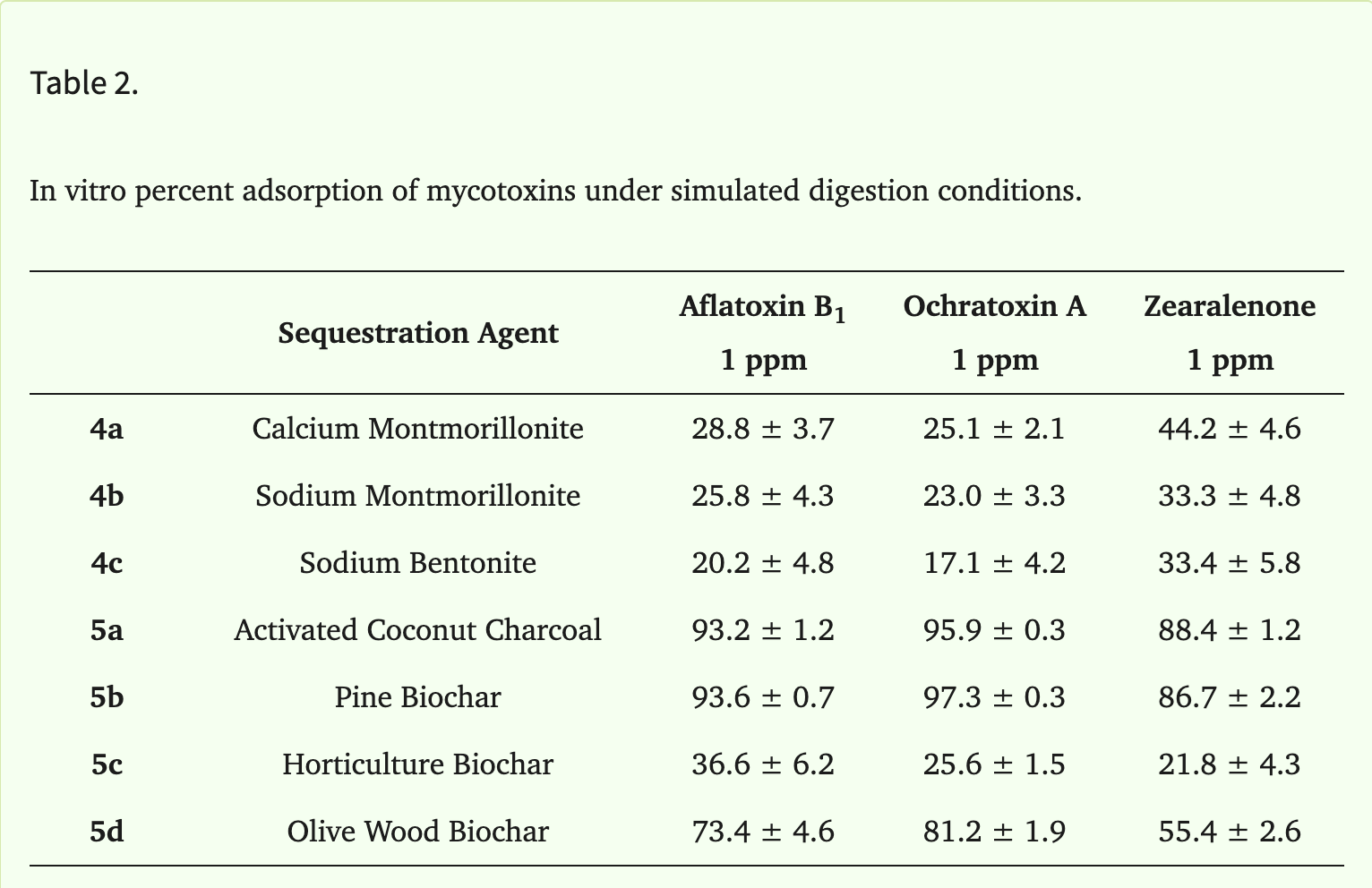
Aflatoxin B1, Ochratoxin A, and Zearalenone: Using several charcoal variations (and clays) like activated coconut charcoal, pine biochar, horticulture biochar, olive wood biochar, inside a simulated gut with varying pH levels and doses of mycotoxin. At a 1ppm mycotoxin dose in the full simulated digestion (stomach then small intestine), they found activated coconut charcoal and pine biochar to be most effective, respectively binding 93.2%/93.6% of aflatoxin B1, 95.9%/97.3% of ochratoxin A, and 88.4%/86.7% of zearalenone. Nobody is going to be taking pine biochar, but everyone has access to coconut charcoal.10
Clay
Aflatoxin: A 3-month human trial in Ghana tested a calcium montmorillonite clay called NovaSil, taken daily before meals, against a placebo. Half of the participants were split into a low dose group of 1.5g/day, and the other 3g/day. The high dose group had a 58% reduction in aflatoxin markers in their blood and urine by month three. There were also no adverse effects attributed to the clay, as blood, liver, and kidney function, and electrolytes were all unaffected, and it didn’t interfere with vitamin A, E, iron, or zinc levels. 11
Aflatoxin: Kenyan participants who were given a form of calcium montmorillonite clay cut their aflatoxin exposure biomarkers roughly in half compared to placebo.12
β-glucan / S. boulardii
Aflatoxins, Ochratoxin, Zearalenone, and Deoxynivalenol: Using a multi-spectrum binder of 70% bentonite clay, 10% humic acid, and 20% beta-glucan-mannan within a simulated gut environment, researchers found this binder removed 98.07% of aflatoxin B1, 93.80% of aflatoxin B2, 90.99% of aflatoxin G1, 93.56% of aflatoxin G2, 81.64% of ochratoxin, 73.45% of zearalenone, and 98.98% of deoxynivalenol. The beta-glucan-mannan boosted binding for ochratoxin, zearalenone, and deoxynivalenol.13
Ochratoxin A: Chicks who were given OTA and S. boulardii had better blood and biochemical markers of liver and kidney damage, which usually accompanies mycotoxin damage, compared to those who were not.14
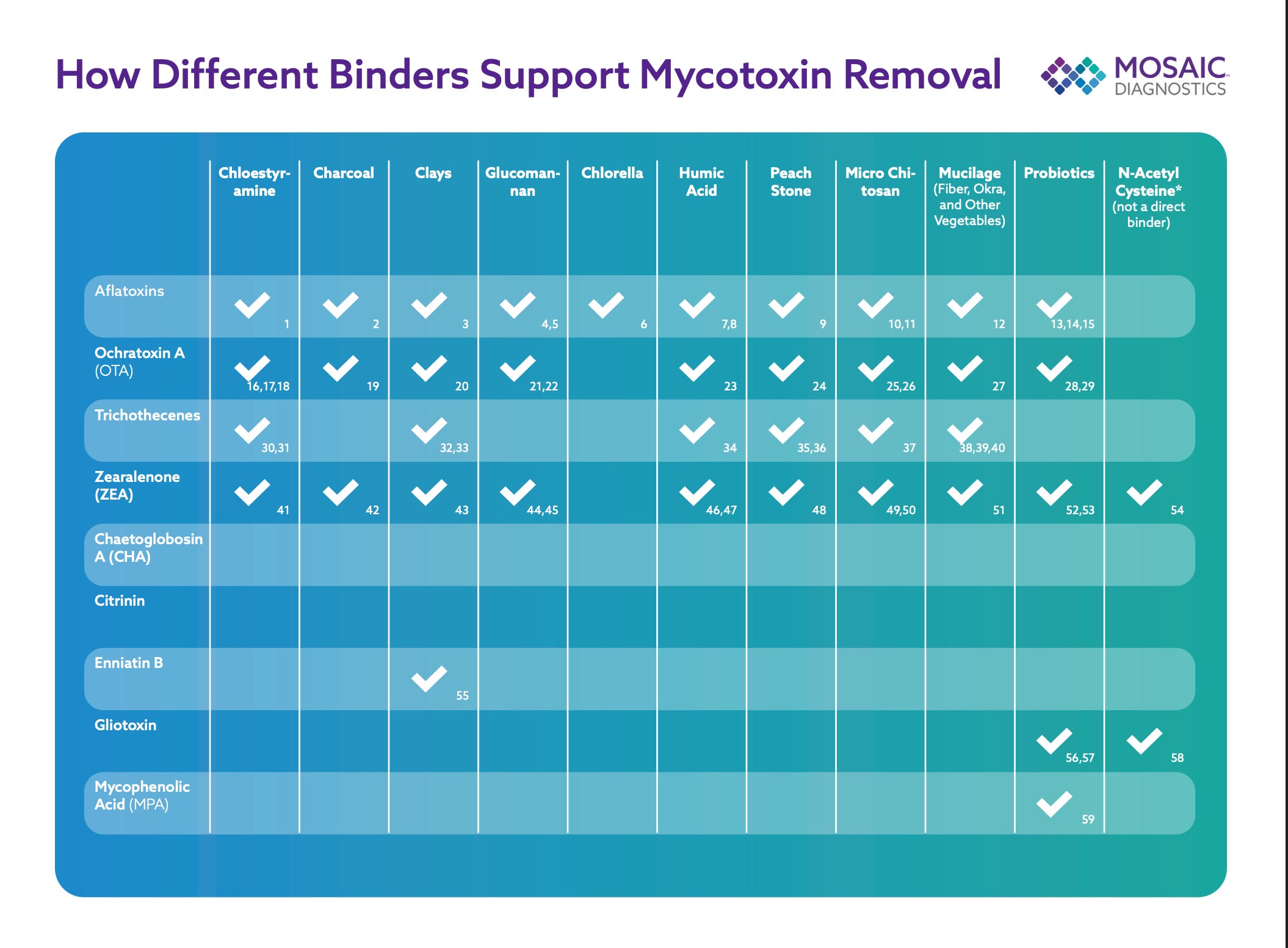
Without going into other binders like chitosan, varying probiotics, and fibers, I will attach a guide from Mosaic Diagnostics, which does some fantastic mycotoxin testing. They compiled tons of research on this topic.15
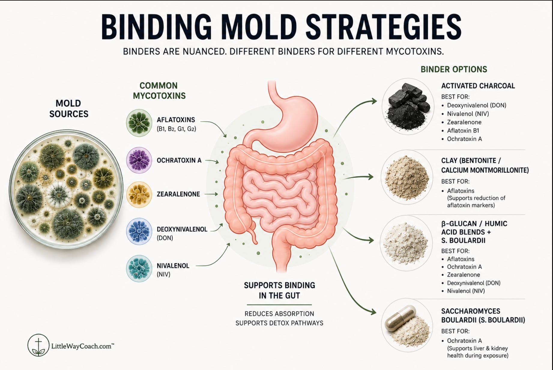
After going deep into the nuances around binding mycotoxins, which frankly, there are many, my opinion is unchanged: a multi-spectrum binder is going to be the most superior strategy.
Extrapolating this out to humans and the real world: a combo of probiotics like S. boulardii, for the cell wall adherence to mycotoxins, activated charcoal, and certain clays would be a great place to start.
While binding might be one of the most critical steps in clearing mycotoxins, it still must be accompanied by other strategies.
This is where I like to build on Shoemaker’s protocols, which in some ways miss the bigger picture, and build on the shoulders of giants like Dr Chris Shade of Quicksilver Scientific.
If you’ve been sick for years, tried everything, and still have no answers, that’s exactly who I work with.
Come read my story, see if it sounds familiar, and book a free consult. It costs you nothing but 20 minutes. Visit littlewaycoach.com to book your consult!

Mycotoxins and Tissue Sequestration
Mycotoxins are not static compounds; rather, they travel the body and sequester themselves into varying tissues.
In 26 human forensic autopsies, from people who died of normal causes, mycotoxins turned up in nearly every tissue tested: fat, liver, kidney, lung, brain, heart.
That same autopsy study, and others, have found fat to be a secondary reservoir for mycotoxin bioaccumulation, meaning, across the mycotoxin spectrum, fat is where the widest range of toxins collect — compared to the liver, which holds the highest concentrations.16
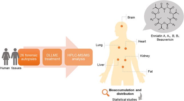
This is also why certain mycotoxins have different effects on the body. And if mycotoxins reside in varying areas of the body, this also means binding, which is done in the gut, is really only one piece of the puzzle when healing from mycotoxins.
In reality, addressing mycotoxins requires a robust framework dedicated to upregulating detox pathways like Nrf2, supporting the detox organs like the liver via bile, addressing the gut dysbiosis that often accompanies a mycotoxin burden, getting ample sunlight, repairing cell membranes, the gatekeeper and brain of the cell, which take a beating from mycotoxins, and using a strategic diet.
Nrf2
Nrf2 is the body’s master detox switch. It’s a protein that flips on certain genes within the cell associated with detoxification and oxidative stress, and its half-life is roughly 15 to 20 minutes.17
Under normal circumstances, Nrf2 is bound and kept in the cell’s cytoplasm, the jelly-like substance within the cell, constantly being degraded and remade, basically on standby until it’s triggered.
Then, when the cell undergoes oxidative stress from the many environmental toxins like mycotoxins, heavy metals, glyphosate, PFAS, endotoxins from the Gram-negative bacteria in the GI tract, or excessive reactive oxygen species coming from dysfunctional mitochondria, Nrf2 stops being degraded and bound in the cytoplasm, and is switched ‘on’.18
It then translocates to the nucleus of the cell, where it binds to a specific DNA sequence called the ARE (Antioxidant Response Element), effectively triggering a cascade affecting over 200 genes associated with protecting the body from these stressors.
Genes that upregulate glutathione synthesis, which squelches ROS and helps conjugate toxins, making them easier to excrete; and genes that build transporters like the multidrug resistance proteins (MRPs), which physically move toxins out of cells.19
The downstream effects of Nrf2 activation are critical for managing environmental toxin loads like mycotoxins, and for managing the body’s natural defense against destructive and excessive oxidative stress (more on these shortly).
Yet Nrf2 and toxins like the mycotoxin ochratoxin A exist on a two-way street. While mycotoxins can activate Nrf2 and the protective genes meant to deal with them, they can also, in substantial loads, inhibit Nrf2 activation entirely.
Researchers reviewing the evidence on this concluded20:
“OTA-induced inhibition of Nrf2 activation and Nrf2 gene transcription together with OTA-induced Nrf2 protein depletion would render the cell defenseless to physiological and compound-induced oxidative stress. This, coupled with the facts that OTA itself induces oxidative stress, and that Nrf2 induction prior to OTA exposure prevents OTA-induced cell death, provides strong evidence that Nrf2 inhibition is the primary mechanism of OTA’s toxicity.”
What does that mean for you and me?
OTA does two damaging things at once. It creates oxidative stress inside the cell, and at the same time it shuts down Nrf2, the system that would have handled that stress. So the harm may not be coming from the oxidative stress by itself; it may be coming from the fact that our natural countermeasure, Nrf2, was shut down.
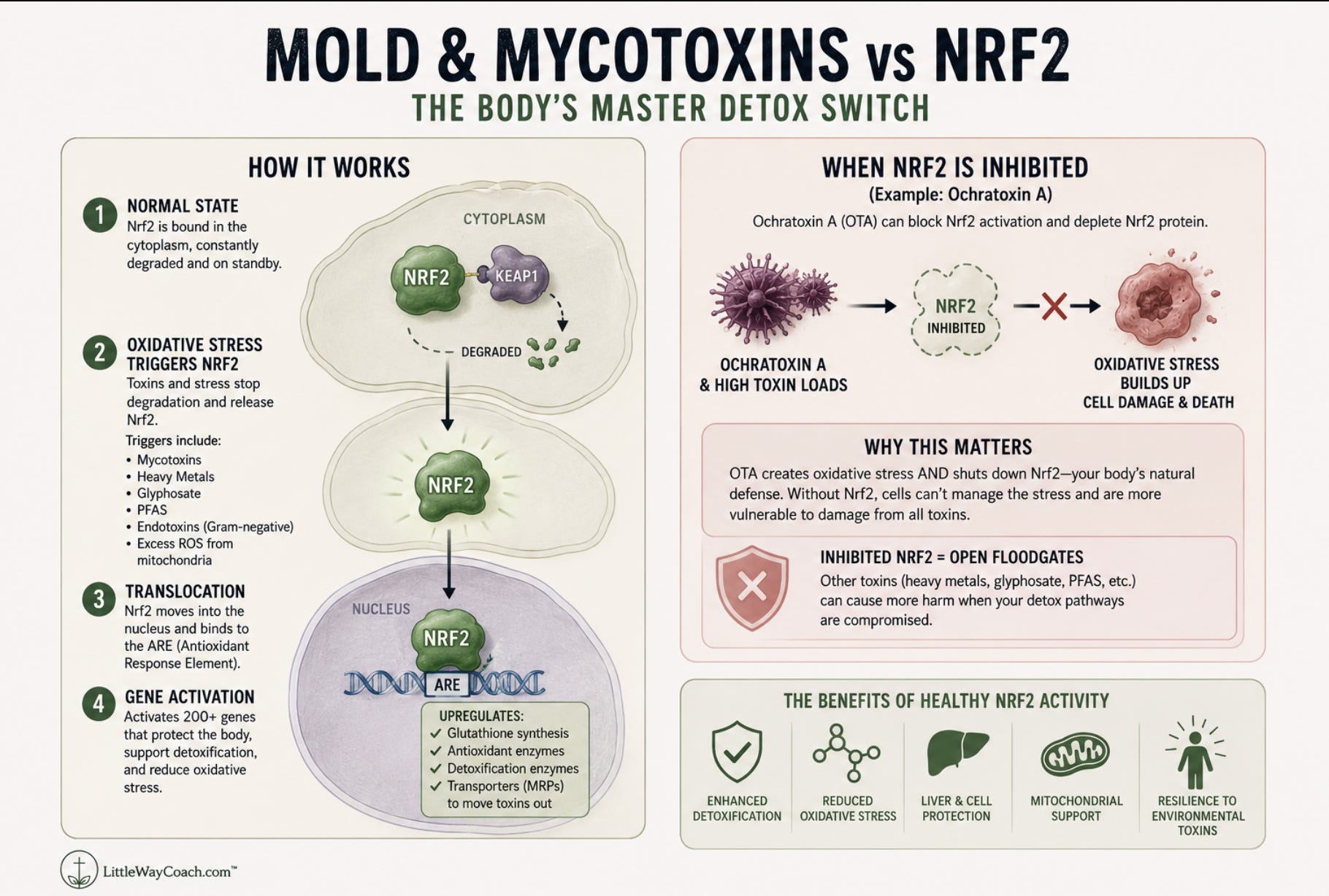
Researchers showed this by inducing Nrf2 in cells before exposing them to OTA. Those cells managed the oxidative stress and survived. The cells that went into the exposure without Nrf2 already active had no countermeasures, got overwhelmed, and died.
This is also why toxic burdens are never just one player. If we are dealing with OTA toxicity, and that’s shutting down our detox master switch, wouldn’t that leave us open to other toxins like heavy metals, glyphosate, and the like?
The floodgates open when our body is overwhelmed, and also why it can take a while to reverse the damage associated with these feedback loops. It’s also another reason you need a robust plan when dealing with mold toxicity, addressing several aspects of the damage.
Glutathione & Mold
Glutathione is a sulfur-based molecule composed of three amino acids: cysteine, glycine, and glutamate. It is one of the body’s most potent antioxidants and plays a critical role in the detoxification process.
It is primarily synthesized in the liver, and while glutathione levels across human tissues range from 0.1 to 10 mM, its highest concentration resides in the liver at up to 10 mM, followed by the spleen, kidney, lens, red blood cells, and white blood cells.21
It’s also responsible for the delicate balance between Th17 dominance and Treg differentiation, meaning it helps determine whether a naive T cell becomes a peacekeeper or a soldier.22
This would help explain its depletion showing up so consistently in autoimmunity, where that same TH17 skew in the adaptive immune system is tightly coupled with low glutathione in autoimmune patients.23
Mitochondria, the powerhouses of the cell, generate their own free radicals as a byproduct of producing energy. But they can’t make their own glutathione — they have to import it from the cytosol using dedicated transporters. Without it, the cell becomes overwhelmed with ROS, which leads to energy failure and, ultimately, apoptosis.24
It’s also responsible for the chelation and conjugation of heavy metals, and serves as a “protective mechanism against possible detrimental effects of the herbicide [glyphosate].”25
God placed one of the body’s master antioxidants and detoxification molecules in places with the highest rates of oxidative stress and toxin processing without mistake, and clearly elevated it as one of the most important molecules in the body.
But sometimes our glutathione levels become overwhelmed; the burden becomes too much. Despite such high concentrations of GSH in the liver, liver cells exposed to OTA mycotoxin showed a 40% reduction in GSH levels after just 24 hours.26
Moreover, revisiting TGF-β, we know high levels of this cytokine push the immune system into “fight or flight” through Th17 dominance and cause tissue damage through fibrogenesis, while depleting glutathione along the way.
It’s been established that “TGF-β increases ROS production and decreases the concentration of glutathione (GSH), the most abundant intracellular free thiol and an important antioxidant, in various types of cells, which mediates many of TGF-β’s fibrogenic effects.”27
Connect some dots with me for a second.
Glutathione and its role in ROS, supporting the mitochondrion’s ability to create energy, its high concentrations in the liver where the body deals with toxins, its role in TH17 dominance, TGF-β, and autoimmunity.
Now connect this to mold and the cascading effect mycotoxins set off:
Mycotoxins like OTA halt Nrf2 and increase ROS
Mycotoxins push the adaptive immune system into fight or flight through increased cytokines like TGF-β
Higher TGF-β pushes more ROS and drives fibrogenesis
Lack of glutathione from steps 1 and 2 means TGF-β’s fibrogenic effects can’t be mediated
Repeat steps 1 through 4, and the damage of mold expands
If glutathione manages so many aspects, and displays such a push-pull relationship with toxins, CIRS, and autoimmunity, shouldn’t we be prioritizing it?
If mold reliably produces the same biochemical picture we find in autoimmune patients, how many people carrying an autoimmune diagnosis have an environmental exposure nobody ever looked for?
I built a free AI tool that knows more about mold than most people ever will. Use it. It’s free. Visit Littlewaycoach.com and ask about mold!

Cell membranes
Cell membranes are one of my favorite topics to cover in my coaching program; it’s rare for clients to know much, if anything, about them.
Cell biologist Bruce Lipton popularized the idea that the membrane, not the nucleus, is the real “brain of the cell.”
The cell membrane determines what comes in, what goes out, which genes we express, how we utilize hormones, how cells communicate with each other, and how we manage toxins, which would make sense of Lipton’s “brain of the cell” terminology.
For example, hormone interactions with the cell membrane occur in three ways: via receptors, transporters, and diffusion.
Peptide hormones like insulin require hormone receptors. They dock onto these receptors, signaling to the cell to act, almost like ringing a doorbell.
Other hormones need transportation. Hormones like thyroid require transporters to carry the hormone from the outside of the cell membrane into the cell.
Compare that to fat-soluble hormones like cortisol and testosterone, which dissolve straight through the lipid bilayer.
Then we have other transporters like the MRPs, the ones Nrf2 builds, which act as pumps embedded in the membrane, pushing toxins out of the cell so they can be excreted.
MRP1 is found in most tissues and is the primary transporter of organic toxins, both conjugated and unconjugated.
Some of these toxins include lipid peroxidation products, herbicides, tobacco-specific nitrosamines, mycotoxins, heavy metals, and natural products and antifolate anti-cancer agents. Our cells need the continual excretion of these toxins from MRP1 transporters in order to prevent a toxic buildup.28
MRP2 pumps are the Phase III step that carries Phase II products out of the cell, specifically targeting fat-soluble toxins and recognizing glutathione conjugates for excretion. They are predominantly found in hepatocytes (liver cells), renal proximal tubular cells (kidney cells), enterocytes (gut cells), and syncytiotrophoblasts of the placenta (placental cells, take note moms!).29
MRP2 pumps are also a driving force behind bile flow, which is a major route for excreting toxins.30
Considering the placement of MRP2 pumps, we can infer the importance of optimizing organs such as the liver, kidneys, and gut for detox. And isn’t it cool how they exist in high loads in the placenta, effectively keeping baby safe from a toxic burden?
So cell membranes are important, clearly, but what about mycotoxins?
Your cell membranes are made of fat — specifically a double layer of fat called a phospholipid bilayer. Between the lipid bilayer and the proteins (like we talked about above), the membrane composition is around 50% lipid and 50% protein by weight.31
Of the phospholipids in the body, one phospholipid named phosphatidylcholine makes up approximately 50% of the phospholipids in the body. Critically, inhibition of PC synthesis in the liver has been linked to fatty liver disease, loss of energy production in the mitochondria, and a higher risk of metabolic diseases like atherosclerosis, insulin resistance, and obesity.32
Regarding mycotoxins, research has linked them to increased lipid peroxidation, distorting the lipids comprising the membrane and disrupting the structure of immune macrophage cells through oxidation.33
Normally, your liver makes and recycles PC two ways: it builds fresh PC from choline and fat, and it recycles PC sent out through bile. Bile itself is recycled at a high rate (up to 95%), so most of that PC gets reused instead of made from scratch.
Fumonisin, a common corn mycotoxin, throws a wrench in this recycling step. It interferes with the pancreatic enzyme that breaks down PC in the gut so it can be reabsorbed, and it damages the pancreas itself, further disrupting that process.
The result? Less PC gets recycled back to the liver, cutting off the steady supply it needs to keep bile flowing and supply the building block for cell membranes.34
You rarely hear about cell membranes, much less in conjunction with mycotoxins, yet it’s clear a healthy cell membrane creates a healthy cell, and a beatdown cell membrane creates a sick cell.
In summary, PC helps rebuild the cell membranes broken down from mycotoxins, and restore the actual bile and detox pathways your body depends on to clear mycotoxins in the first place.35
Always be PC maxxing!
Wrapping Up
As much as I wanted to keep writing this one, and expanding even further on strategies, it would have taken me another two weeks, and this article has already taken me two and a half as it is. My brain is fried wrapping this one up.
The amount of research I went through was astonishing, and it taught me a lot while solidifying my stance: mold requires a deep and robust framework.
The next one will get into more of the strategies: gut health, liver health, sun exposure, and immune system function.
This article is for educational purposes only and is not medical advice. I am a health coach, not a physician, and nothing here is intended to diagnose, treat, cure, or prevent any disease. Always work with a qualified healthcare provider before making changes to your health protocol, especially if you are taking medications or managing a diagnosed condition.
https://www.ncbi.nlm.nih.gov/books/NBK143943/ https://pmc.ncbi.nlm.nih.gov/articles/PMC3114807/
https://www.survivingmold.com/docs/12_STEP_SHOEMAKER_PROTOCOL_FOR_CIRS.PDF
https://pubmed.ncbi.nlm.nih.gov/18612139/ https://pubmed.ncbi.nlm.nih.gov/28103316/
https://pmc.ncbi.nlm.nih.gov/articles/PMC10468540/
https://www.survivingmold.com/docs/VIP_published_3_2013.pdf
https://www.survivingmold.com/about/ritchie-shoemaker-m-d
https://pubmed.ncbi.nlm.nih.gov/9482354/
https://www.ncbi.nlm.nih.gov/pmc/articles/PMC4210882/
https://pubmed.ncbi.nlm.nih.gov/15046828/
https://pmc.ncbi.nlm.nih.gov/articles/PMC10649945/#sec3-animals-13-03311
https://pubmed.ncbi.nlm.nih.gov/18478481/
https://pmc.ncbi.nlm.nih.gov/articles/PMC5561728/
https://www.nature.com/articles/s41598-025-13249-z
https://vetsci.org/DOIx.php?id=10.4142/jvs.2004.5.4.359
https://mosaicdx.com/wp-content/uploads/2026/04/Binders-Chart.pdf
https://pmc.ncbi.nlm.nih.gov/articles/PMC10761373/#Sec11
https://www.frontiersin.org/journals/toxicology/articles/10.3389/ftox.2026.1870680/full
https://www.spandidos-publications.com/10.3892/ijmm.2014.1696#b32-ijmm-33-05-1348
https://townsendletter.com/a-push-catch-system-that-enables-effective-detoxification/
https://portlandpress.com/biochemj/article/395/3/599/44215/Role-of-Nrf2-in-the-regulation-of-the-Mrp2-ABCC2
https://pubmed.ncbi.nlm.nih.gov/17668877/
https://www.ncbi.nlm.nih.gov/pmc/articles/PMC3920267/
https://www.sciencedirect.com/science/article/abs/pii/S0009898103002006
https://elifesciences.org/articles/36158
https://pubmed.ncbi.nlm.nih.gov/19393193/
https://pmc.ncbi.nlm.nih.gov/articles/PMC2821140/#s012
https://pubmed.ncbi.nlm.nih.gov/23044091/
https://pmc.ncbi.nlm.nih.gov/articles/PMC8068875/#abstract1
https://pmc.ncbi.nlm.nih.gov/articles/PMC2818240/
https://pubmed.ncbi.nlm.nih.gov/11557126/
https://pubmed.ncbi.nlm.nih.gov/16863439/
https://pubmed.ncbi.nlm.nih.gov/16847695/
https://www.ncbi.nlm.nih.gov/books/NBK9898/
https://www.sciencedirect.com/science/article/pii/S0005273617301220
https://www.sciencedirect.com/science/chapter/edited-volume/abs/pii/B9780128190920000200?via%3Dihub
https://www.sciencedirect.com/science/article/pii/S0300483X24001410
https://www.sciencedirect.com/science/article/abs/pii/S0378427401004763
https://bodybio.com/blogs/blog/how-to-protect-yourself-against-mold
https://pmc.ncbi.nlm.nih.gov/articles/PMC6989179/
https://www.sciencedirect.com/science/article/pii/S0005273613004070 https://www.ncbi.nlm.nih.gov/books/NBK470209/